
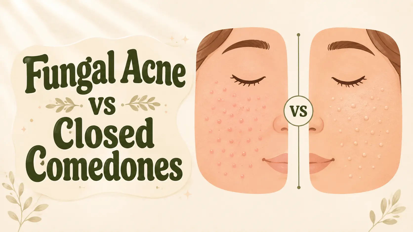
“Fungal acne” usually means Malassezia folliculitis, which is not acne. It tends to cause small follicular papules and pustules that look fairly similar to one another, often itch, and usually appear on the chest, upper back, shoulders or hairline. Closed comedones are true acne lesions: pores plugged with sebum and keratin, seen as flesh-coloured or whitish bumps, commonly mixed with blackheads and other acne lesions. A key textbook distinction is that comedones are absent from Malassezia folliculitis lesions themselves. However, Malassezia folliculitis and acne can coexist, so a person with MF may also have separate acne comedones. Appearance alone cannot reliably diagnose every case.1,2,4,10,13
The phrase “fungal acne” is an informal term widely used online and on social media. Dermatology literature calls the condition Malassezia folliculitis (formerly Pityrosporum folliculitis). Malassezia yeasts normally live on human skin; disease occurs when follicular overgrowth becomes associated with inflammation and symptoms.1,2,3,14
Acne vulgaris, bacterial folliculitis and other acneiform or follicular eruptions can resemble Malassezia folliculitis. Repeatedly rotating antifungals, antibiotics and exfoliants without knowing the diagnosis can delay appropriate care or irritate the skin.2,3,14
In this guide
- Fungal acne vs closed comedones at a glance
- What Malassezia folliculitis actually is
- What closed comedones are
- Appearance, itching and location
- How dermatologists diagnose Malassezia folliculitis
- Can you have both?
- Internet claims: what is evidence and what is not
- Treatment of Malassezia folliculitis
- Treatment of closed comedones
- When to see a dermatologist
- Bottom line
- Frequently asked questions
Fungal acne vs closed comedones: the comparison
| Feature | Malassezia folliculitis | Closed comedones |
|---|---|---|
| What it is | Inflammation of hair follicles associated with overgrowth of Malassezia yeast. | A clinically non-inflammatory acne lesion caused by follicular plugging. |
| Typical lesion | Small follicular papules and pustules. | Flesh-coloured or whitish closed bumps/whiteheads. |
| Comedones | Absent from MF lesions. Separate acne comedones can coexist if both conditions are present.1,2,4,10 | Present by definition. |
| Uniformity | Often described as monomorphic: lesions are similar in size and stage. | May coexist with blackheads, inflamed pimples and lesions of different sizes/stages. |
| Itch | Common, but not universal. One 26-patient series reported itching in 70%.7 | Not usually a defining symptom. |
| Common sites | Upper chest, back, shoulders/upper arms; face, hairline, neck can also be involved.3,4 | Face is common; chest and back can also develop comedonal acne. |
| Typical context | Heat/humidity, sweating, occlusion, antibiotic exposure or immunosuppression can increase suspicion.1,4 | Acne biology includes follicular hyperkeratinisation, sebum and inflammation; hormones and genetics matter. |
| Response to antibiotics | Often fails to improve; antibiotic exposure is a recognized predisposing context for Malassezia folliculitis.3,7,14 | Antibiotics can help inflammatory acne when appropriately used, but they are not the main treatment for isolated closed comedones. |
| Confirmation | Clinical pattern plus demonstration of yeast in follicular material by microscopy/staining; biopsy can help difficult cases.1,2,5 | Usually a clinical acne diagnosis. |
What “fungal acne” actually is
Malassezia is part of the normal human skin mycobiome. These yeasts are strongly associated with lipid-rich skin sites. Most Malassezia species are lipid-dependent; genomic and growth studies show loss of cytosolic fatty-acid synthase capacity and dependence on environmental lipids or fatty acids for growth.2,12
That biology does not mean that simply detecting Malassezia on skin proves disease. Healthy skin also carries the organism. In Malassezia folliculitis, yeast is found in the follicular environment together with a compatible clinical eruption and inflammation. This is why expert recommendations focus on the combination of clinical features and positive diagnostic findings, not a cosmetic-ingredient list alone.1,2
Calling this “fungal acne” is convenient for search, but it can also create confusion: acne is a different follicular disorder. Malassezia folliculitis is a form of folliculitis.
What closed comedones are
Closed comedones are plugged follicles in acne vulgaris. The opening is not visibly open to the surface the way a blackhead is, so they appear as small skin-coloured or pale bumps. They can remain non-inflammatory or later become inflamed.
Modern acne guidelines strongly recommend topical retinoids because they help normalise follicular keratinisation and unclog pores. Salicylic acid is also recommended as a topical option, although the strength of evidence differs across therapies.8,9
If your main problem is acne rather than folliculitis, see our evidence-based acne treatment guide and our salicylic acid vs benzoyl peroxide comparison.
Can you tell them apart by appearance?
1. Monomorphic vs polymorphic lesions
Dermatology sources repeatedly describe Malassezia folliculitis as monomorphic: many small papules/pustules at a similar stage. Acne vulgaris is more often polymorphic, with a mixture of closed comedones, open comedones, inflammatory papules, pustules and sometimes deeper lesions.2,3,4
But “all my bumps look the same” is not a diagnostic test. Similar-looking bumps occur in several follicular disorders.
2. Are comedones present?
This is a key textbook distinction. True comedones argue for acne. Expert reviews and clinical references specifically note that comedones are not a feature of Malassezia folliculitis.1,2,4
3. Is it itchy?
Itch increases suspicion for Malassezia folliculitis, but the popular claim that it is always itchy is wrong. In one clinical series, 70% of patients reported itching—meaning a substantial minority did not.7 Itch can also occur with dermatitis, irritation, heat rash and other conditions.
4. Where are the bumps?
Malassezia folliculitis classically affects the upper trunk—especially chest and back—and may involve shoulders, posterior arms, forehead/hairline, chin and neck.3,4 Closed comedonal acne is particularly common on the face but can occur on the trunk too. Location helps; it does not prove the diagnosis.
Uniform + itchy + upper trunk + no comedones is a useful pattern of suspicion, not a home laboratory test.
Stylish & Healthy evidence noteHow dermatologists diagnose Malassezia folliculitis
A clinician can suspect the condition from the lesion pattern, distribution, itch and history—especially when an acne-like eruption does not respond to standard acne therapy. But because Malassezia normally lives on skin, diagnosis is more meaningful when sampling comes from the follicular contents of a compatible lesion.1,2
Direct microscopy and stains
Potassium hydroxide (KOH) preparation and other direct microscopy/staining techniques can demonstrate yeast. In a study of 264 patients with folliculitis, 49 were diagnosed with Malassezia folliculitis; among those MF cases, May-Grünwald-Giemsa cytology was positive in 100% and KOH preparation in 81.6%. The exact performance of a test depends on sampling and technique, so these figures should not be treated as universal sensitivity estimates.5
Biopsy
Histopathology can be useful when the diagnosis remains uncertain or when another form of folliculitis must be excluded. Reviews describe follicular dilation/inflammation with Malassezia organisms within affected follicles.2
Culture is awkward
Malassezia is lipid-dependent and can be difficult to culture on routine media. A negative routine fungal culture therefore does not necessarily settle the question; specialist methods may be needed.2
What about a treatment trial?
Rapid improvement with antifungal therapy can support a suspected diagnosis, but using treatment response as the only “test” is imperfect. Rashes fluctuate, treatments can have nonspecific effects, and unnecessary medication carries risks.1,2
Can you have Malassezia folliculitis and acne at the same time?
Yes. The conditions are not mutually exclusive. Reviews report coexistence, and a study specifically examined Malassezia folliculitis among patients who already had papulopustular or comedonal acne.3,10
This matters because a person may have obvious blackheads and closed comedones plus a separate crop of itchy monomorphic follicular bumps. Treating only one process can make the overall picture look “treatment-resistant.”
Four internet claims that need more nuance
“Fungal acne is always itchy.”
Too absolute. Pruritus is common and diagnostically useful, but clinical series include non-itchy cases.7
“Uniform bumps prove fungal acne.”
No. Monomorphic follicular papules/pustules increase suspicion, but several acneiform and follicular eruptions can look uniform.
“Certain skincare ingredients feed fungal acne.”
Biologically plausible in a laboratory sense, clinically oversimplified. Malassezia is lipid-dependent and can hydrolyse/use some fatty acids and esters, but laboratory substrate utilisation does not establish that a specific cosmetic ingredient will trigger folliculitis on human skin at real-world concentrations.11,12
“A fungal-acne-safe ingredient checker can tell you what to buy.”
In our literature search, last checked August 9, 2026, we did not identify a clinically validated universal checker. Laboratory studies show that Malassezia is lipid-dependent and can utilise some fatty acids or esters, but those studies do not establish that an ingredient blacklist predicts MF flares in people.11,12
So do oils and occlusive products matter at all?
They can matter, but the evidence is broader than internet blacklists. Clinical references list occlusion, sweating, hot/humid conditions and some emollient/sunscreen use as possible risk factors or flare contexts.3,4 That supports paying attention to your actual clinical pattern and product experience. It does not justify declaring dozens of individual esters universally forbidden.
How Malassezia folliculitis is treated
Antifungal therapy is the core treatment. European expert recommendations and reviews discuss topical azoles for milder disease and systemic azoles for more extensive or difficult cases, with the exact choice depending on the patient and local prescribing practice.1,2
Topical antifungals
Ketoconazole and other topical antifungals are used clinically. In a small, non-randomized Japanese series of 44 patients, 37 received topical 2% ketoconazole and 7 received oral itraconazole until lesions flattened. Mean time to improvement was shorter in the oral group (14 ± 4 days) than the topical group (27 ± 16 days), but the groups were small and treatment choice depended on severity and patient preference, so this should not be read as a definitive head-to-head trial.6
Oral antifungals
Itraconazole and fluconazole have published evidence in Malassezia folliculitis, but oral azoles are prescription medications with meaningful contraindications and drug-interaction considerations. They should not be self-prescribed from an online diagnosis. Expert recommendations specifically tailor systemic therapy according to immune status and liver function.1,2
Recurrence
Relapse is common. In the 26-patient series, recurrence occurred within months after treatment stopped, and reviews discuss intermittent topical maintenance for recurrent disease.2,7
Why acne treatment may seem to “make fungal acne worse”
Antibiotic exposure is repeatedly reported as a predisposing context for Malassezia folliculitis.1,3,14 This does not mean every acne ingredient worsens Malassezia. Retinoids, salicylic acid and benzoyl peroxide have different mechanisms from antibiotics.
How closed comedones are treated
Closed comedones need an acne strategy, not an antifungal strategy. The 2024 AAD guideline strongly recommends topical retinoids such as adapalene, tretinoin, tazarotene and trifarotene as acne therapies, and also recommends benzoyl peroxide and several other topical options. Salicylic acid and azelaic acid receive recommendations as well.8,9
- Topical retinoid: usually the backbone for comedonal acne because it targets abnormal follicular keratinisation.
- Salicylic acid: can help unclog pores and exfoliate within an acne routine.
- Azelaic acid: can be useful when acne and post-acne pigmentation overlap.
- Moisturizer + sunscreen: support tolerability so effective acne treatment is easier to continue.
Do not respond to every new bump by stacking more acids. Over-irritation can create redness, burning and rough texture that can obscure the original clinical picture.9 Our damaged skin barrier guide explains those warning signs.
When to see a dermatologist
- You have persistent itchy follicular bumps that have not responded to a sensible acne routine.
- You have been using antibiotics and an acne-like eruption is becoming more uniform or itchy.
- The rash is widespread across the chest/back, keeps recurring, or is leaving marks.
- You are immunosuppressed, have significant medical conditions, or are considering oral antifungal treatment.
- You cannot tell whether the bumps are acne, folliculitis, dermatitis, milia or another condition.
- You develop painful nodules, rapidly spreading redness, fever, drainage or other signs of significant infection.
The bottom line
Malassezia folliculitis and closed comedones can look similar in a phone photo, but they are not the same disease. The classic MF pattern is itchy, monomorphic follicular papules/pustules on sebaceous areas such as the upper trunk, with no comedones. Closed comedones are acne plugs and often live alongside blackheads or inflammatory acne.
The most important correction to social-media advice is this: you cannot reliably diagnose “fungal acne” from bump uniformity or an ingredient checker. When the diagnosis matters, dermatology examination and follicular microscopy are much more defensible than guessing from a list of “safe” and “unsafe” esters.
Frequently asked questions
Is fungal acne really fungus?
The common term refers to Malassezia folliculitis, which involves yeast of the genus Malassezia. Yeasts are fungi, but the condition is folliculitis—not acne.1,2
Does fungal acne always itch?
No. Itch is common, but not universal. One clinical series reported pruritus in 70% of patients.7
Can fungal acne have whiteheads?
Malassezia folliculitis can form small pustules that may look white-tipped, but true comedones are not a typical feature. Closed comedones point toward acne vulgaris.1,4
Can I have both fungal acne and closed comedones?
Yes. Malassezia folliculitis can coexist with acne vulgaris, which is one reason mixed eruptions can be confusing.3,10
Does salicylic acid treat fungal acne?
Salicylic acid is an acne treatment and is recommended in acne guidance, but it is not the primary antifungal treatment for confirmed Malassezia folliculitis.1,8,14
Are “fungal acne safe” products scientifically proven?
In our literature search, last checked August 9, 2026, we did not identify a clinically validated universal ingredient-checker system. Laboratory studies show that Malassezia is lipid-dependent, but translating lipid utilisation directly into a blacklist of cosmetic ingredients goes beyond what those studies establish in people.11,12
References
- Henning MAS, et al. Position statement: Recommendations on the diagnosis and treatment of Malassezia folliculitis. J Eur Acad Dermatol Venereol. 2023;37(7):1268-1275.
- Saunte DML, Gaitanis G, Hay RJ. Malassezia-Associated Skin Diseases, the Use of Diagnostics and Treatment. Front Cell Infect Microbiol. 2020;10:112.
- Rubenstein RM, Malerich SA. Malassezia (Pityrosporum) Folliculitis. J Clin Aesthet Dermatol. 2014;7(3):37-41.
- DermNet. Malassezia (pityrosporum) folliculitis. Clinical features, diagnosis and treatment.
- Durdu M, Güran M, Ilkit M. Epidemiological characteristics of Malassezia folliculitis and use of the May-Grünwald-Giemsa stain to diagnose the infection. Diagn Microbiol Infect Dis. 2013;76(4):450-457.
- Suzuki C, et al. Treatment Outcomes for Malassezia Folliculitis in the Dermatology Department of a University Hospital in Japan. Med Mycol J. 2016;57:E63-E66.
- Lévy A, Feuilhade de Chauvin M, Dubertret L, Morel P, Flageul B. Malassezia folliculitis: characteristics and therapeutic response in 26 patients. Ann Dermatol Venereol. 2007;134(11):823-828. PMID: 18033060.
- Reynolds RV, Yeung H, Cheng CE, et al. Guidelines of care for the management of acne vulgaris. J Am Acad Dermatol. 2024;90(5):1006.e1-1006.e30. doi:10.1016/j.jaad.2023.12.017. PMID: 38300170.
- American Academy of Dermatology. Updated guidelines for the management of acne. 2024.
- Pürnak S, Durdu M, Tekindal MA, Güleç AT, Seçkin D. The Prevalence of Malassezia Folliculitis in Patients with Papulopustular/Comedonal Acne, and Their Response to Antifungal Treatment. SKINmed. 2018;16(2):99-104. PMID: 29911526.
- Mayser P, et al. Hydrolysis of fatty acid esters by Malassezia furfur: different utilization depending on alcohol moiety. Acta Derm Venereol. 1995;75(2):105-109.
- Liebregts J, et al. Lipid-dependent growth of Malassezia spp. in defined medium with single fatty acids. FEMS Yeast Res. 2025;25:foaf043.
- Chalupczak NV, Lipner SR. Malassezia Folliculitis: An Underdiagnosed Mimicker of Acneiform Eruptions. J Fungi (Basel). 2025;11(9):662. doi:10.3390/jof11090662. PMID: 41003208; PMCID: PMC12471122.
- Draelos ZD, Barbieri JS, Tanghetti EA, et al. Malassezia Folliculitis Presentation, Diagnosis, and Treatment: A Review of “Fungal Acne”. J Drugs Dermatol. 2026;25(5):427-434. doi:10.36849/JDD.9751. PMID: 42081639.
Medical disclaimer: This article is for education only and is not a diagnosis or a substitute for care from a qualified health professional. Skin eruptions that are persistent, painful, rapidly worsening or associated with systemic symptoms should be assessed clinically.
Evidence first. Hype second.